
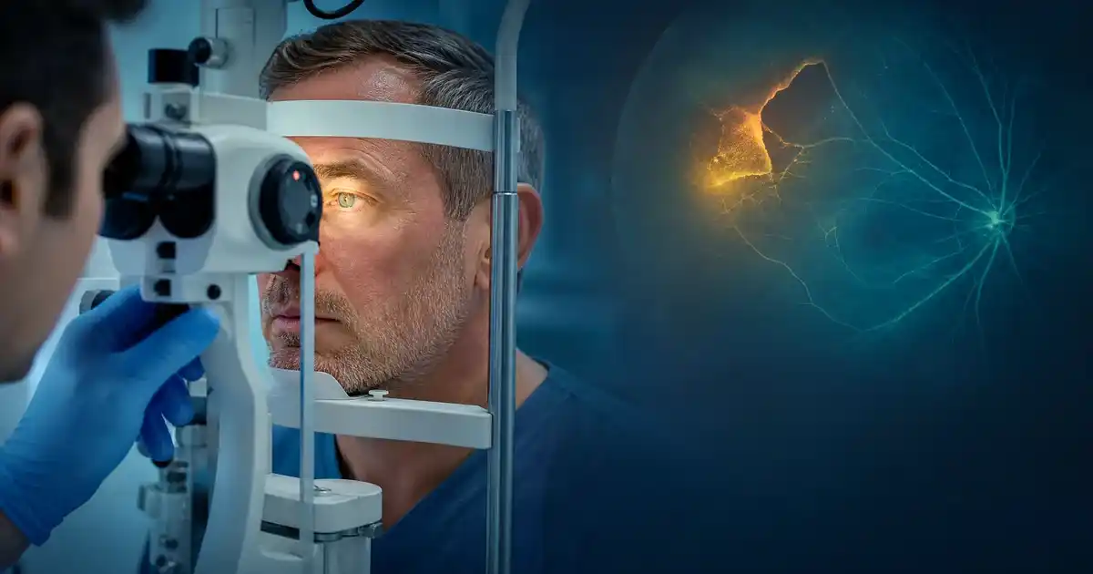
Retinal Tear: How Is It Diagnosed and Treated Before Vision Loss?
A retinal tear may cause sudden floaters or flashes and requires timely diagnosis and appropriate treatment before it progresses to retinal detachment and vision loss.
A retinal tear is an eye condition that requires prompt attention because, in some cases, a tear can allow fluid to pass underneath the retina and contribute to retinal detachment. A retinal tear may not cause pain, so sudden changes in vision, especially a new increase in floaters or flashes of light, can be important warning signs.
The appropriate approach to a retinal tear depends on its location, size, appearance, and whether there are signs that the retina has already started to detach. Many retinal tears can be treated before retinal detachment develops, commonly with laser treatment or cryotherapy when appropriate. This article by Dr. Khalil Ibrahim Hijazi Consultant & Ophthalmic Surgeon explains the main retinal tear causes, symptoms, diagnostic methods, treatment options, and the relationship between retinal tears, retinal detachment, and vision loss.
What Is a Retinal Tear and What Causes It?
The retina is the light-sensitive tissue lining the back of the eye. A retinal tear occurs when a break or split develops in the retinal tissue. It can be associated with changes in the vitreous gel inside the eye, particularly when the vitreous pulls on the retina. A retinal tear does not always mean that retinal detachment has already occurred, but it requires careful assessment because some tears can progress.
Retinal Tear
A retinal tear is a break in the retinal tissue caused by traction between the vitreous and the retina. It may develop suddenly, particularly during or after a posterior vitreous separation. Some patients experience flashes or new floaters, while other retinal tears may initially produce few or no noticeable symptoms.
Early detection is important because a retinal tear may be treated before retinal detachment develops. The ophthalmologist assesses the location and characteristics of the tear and examines the surrounding retina before deciding whether laser treatment, cryotherapy, or observation is appropriate.
You can also learn more about warning signs associated with retinal detachment in “Retinal Detachment: Warning Signs That Should Not Be Ignored”.
Retinal Tear Causes
Common retinal tear causes include age-related changes in the vitreous, which can pull on the retina as the vitreous separates from it. In some individuals, this traction can create a retinal break.
Other risk factors include significant myopia, previous eye surgery, serious eye trauma, a personal history of retinal tear or detachment in the other eye, a family history of retinal detachment, and areas of retinal weakness identified during an eye examination.
Having a risk factor does not mean that a retinal tear will definitely occur. However, people at higher risk should be particularly attentive to sudden visual changes and follow the eye examinations recommended by their ophthalmologist.
Is a Retinal Tear Dangerous?
Whether a retinal tear is dangerous depends on its characteristics and the condition of the surrounding retina. A retinal tear can become serious if it progresses to retinal detachment, because fluid may pass through the tear and lift the retina away from its normal position.
However, a retinal tear does not automatically mean that permanent vision loss will occur. When detected before retinal detachment, many retinal tears can be treated with laser or cryotherapy to create a barrier around the tear and reduce the risk of progression.
Symptoms and Signs of a Retinal Tear
Retinal tear symptoms may appear suddenly and can occur when the vitreous pulls on the retina or when there is bleeding inside the eye. New visual symptoms should be evaluated because symptoms alone cannot confirm whether a retinal tear is present.
Retinal Tear Symptoms
Common retinal tear symptoms include sudden flashes of light, a sudden increase in floaters, or new spots, lines, or cobweb-like shapes moving across the visual field. Some people may also notice blurred vision or changes in peripheral vision.
A retinal tear is often painless, so the absence of pain does not rule it out. At the same time, flashes and floaters do not always mean that a retinal tear is present, as they can have other causes. New or sudden symptoms should therefore be assessed by an eye specialist.
Signs of Retinal Tear
Important signs of retinal tear include a sudden increase in floaters, repeated flashes of light, a shadow in the field of vision, or a sudden change in vision.
A dark curtain or expanding shadow is particularly concerning because it may indicate retinal detachment rather than an isolated retinal tear. Such symptoms require urgent ophthalmic assessment rather than waiting for them to disappear.
Eye Floaters and Retinal Tear
Eye floaters and retinal tear can be associated when floaters suddenly appear or increase significantly over a short period. Floaters may look like small dots, lines, strands, or cobwebs moving across the field of vision.
Long-standing, stable floaters do not necessarily indicate a retinal tear. The greater concern is a sudden increase in floaters, especially when they occur together with flashes or changes in vision. A dilated retinal examination may be needed to exclude a retinal tear or retinal detachment.
Eye Flashes and Retinal Tear
Eye flashes and retinal tear may be connected to traction between the vitreous and retina. Patients may describe flashes as brief streaks or bursts of light, often noticed in the peripheral vision or in dim environments.
Not every flash indicates a retinal tear. However, sudden flashes, especially when accompanied by new floaters, a shadow, or changes in the visual field, should be evaluated promptly.
How Is a Retinal Tear Diagnosed?
Retinal tear diagnosis depends on a detailed retinal examination, usually after dilating the pupil. The ophthalmologist examines the retina carefully, including the peripheral areas where some retinal tears can occur.
Retinal Tear Diagnosis
Retinal tear diagnosis begins with a detailed discussion of the symptoms. The doctor may ask when the flashes or floaters started, whether they appeared suddenly, and whether there has been previous eye trauma, surgery, high myopia, or a history of retinal problems.
A dilated retinal examination is then performed. A retinal specialist may use specialized lenses and examination techniques, including scleral depression when appropriate, to inspect the peripheral retina. If the view of the retina is limited by bleeding or another opacity, ocular ultrasound may be useful in selected cases.
For more information about preventive eye assessment, see “Comprehensive Eye Examinations for Early Detection of Vision Disorders”.
Retinal Examination
A retinal examination usually involves dilating the pupil with eye drops and examining the fundus using specialized lenses and instruments. The doctor looks for retinal tears, holes, weak areas, bleeding, retinal detachment, or fluid beneath the retina.
The examination can be important even when central visual acuity appears normal because a peripheral retinal tear may not immediately affect central vision.
Additional imaging may be considered when the retina cannot be adequately visualized, but the appropriate diagnostic method depends on the clinical findings.
Sudden Retinal Tear
A sudden retinal tear requires prompt assessment, particularly when accompanied by numerous new floaters, flashes, or changes in the visual field. The purpose of rapid diagnosis is to identify the tear before fluid passes through it and contributes to retinal detachment.
A curtain-like shadow, expanding dark area, or sudden reduction in vision should be treated as an urgent warning sign rather than something to monitor at home.
Treatment of a Retinal Tear Before Detachment
Treatment of a retinal tear before detachment depends on the tear's location, size, appearance, and associated retinal findings. Some tears can be treated with laser or cryotherapy, while a tear associated with retinal detachment may require a surgical approach.
Retinal Tear Treatment
The goal of retinal tear treatment is to create a secure barrier around the tear and reduce the risk of fluid passing underneath the retina. Laser retinopexy is commonly used for suitable retinal tears, while cryotherapy may be considered in selected situations.
Not every retinal tear requires immediate treatment. Some low-risk, asymptomatic tears may be monitored rather than treated, depending on their appearance and the individual patient's retinal findings.
Laser Treatment for Retinal Tear
Laser treatment for retinal tear involves applying laser spots around the retinal tear to create an adhesion that helps stabilize the retina and limit the progression of the break.
The procedure may be performed using topical or local anesthesia and is generally less invasive than surgery inside the eye. Temporary glare or blurred vision may occur after treatment, and existing floaters may remain because laser treatment does not remove them. Follow-up is important to confirm that the treatment has produced the intended barrier and to monitor for additional retinal tears.
Treatment of Retinal Tear Before Detachment
The importance of treatment of retinal tear before detachment is that early management can reduce the likelihood of progression to retinal detachment, which may require more complex surgery.
Condition | Typical assessment | Possible management |
|---|---|---|
Low-risk retinal tear | Detailed retinal examination | Observation when appropriate |
Retinal tear requiring sealing | Dilated retinal assessment | Laser or cryotherapy |
Retinal tear with early retinal changes | Urgent retinal assessment | Management based on findings |
Retinal detachment | Surgical retinal evaluation | Vitrectomy, scleral buckle, pneumatic retinopexy, or another appropriate procedure |
A retinal tear and retinal detachment are not the same condition. A tear is a break in the retina, while retinal detachment occurs when the retina separates from its normal position. A tear can allow fluid to pass underneath the retina and contribute to detachment in some cases.
The Relationship Between Retinal Tear and Vision Loss
Not every retinal tear causes vision loss. The main concern is that an untreated or progressive tear can contribute to retinal detachment, which may threaten vision, particularly when the central retina becomes involved.
Retinal Tear and Retinal Detachment
Retinal tear and retinal detachment represent different stages of retinal disease. A tear is an opening in the retinal tissue, whereas detachment occurs when the retina separates from the tissue beneath it. In some cases, fluid can pass through a retinal tear and lift the retina from its normal position.
This is why new flashes, a sudden increase in floaters, or a curtain-like shadow should not be ignored. Early assessment may allow an appropriate retinal tear to be treated before detachment occurs.
Retinal Tear Surgery
The term retinal tear surgery does not necessarily mean that every retinal tear requires surgery inside the eye. If the tear is localized and the retina remains attached, laser treatment or cryotherapy may be sufficient.
If retinal detachment has developed, however, surgery may be required. Depending on the condition, procedures may include vitrectomy, scleral buckling, or pneumatic retinopexy. The choice depends on the location and extent of the detachment, the retinal breaks, and the condition of the vitreous and retina.
Prevention of Retinal Tear
Not all retinal tears can be prevented because some are related to natural changes in the vitreous and aging. However, early recognition of warning signs can help prevent delays in diagnosis.
People should seek an eye examination when they experience a sudden increase in floaters, new flashes, a shadow, a curtain-like area, or sudden changes in vision. Individuals with known risk factors should also follow the retinal monitoring schedule recommended by their ophthalmologist.
For more information about retinal assessment, see “Fundus Imaging: When Is It Recommended and Why Is It Important?” and “Diabetic Retinopathy: Symptoms and Treatment Options to Protect Vision”.
Information about general eye protection is also available in “How to Protect Your Eyes from UV Radiation Throughout the Year”.
Conclusion
A retinal tear requires careful and timely assessment, especially when it appears with new flashes, sudden floaters, or changes in the visual field. A retinal tear does not automatically mean permanent vision loss, and many tears can be managed before they progress to retinal detachment. Treatment depends on the characteristics of the tear and may include laser treatment, cryotherapy, or surgery when retinal detachment has already developed.
Understanding retinal tear symptoms and recognizing warning signs can help patients seek appropriate evaluation without delay. Detailed retinal examination and follow-up remain important for protecting vision and detecting any new retinal changes. The appropriate diagnostic and treatment plan can be discussed with Dr. Khalil Ibrahim Hijazi Consultant & Ophthalmic Surgeon based on the individual examination findings.
Frequently Asked Questions
Related Articles
Corneal Transplant: Suitable Cases and the Success Rate of the Procedure
This article explains corneal transplant indications, suitable cases, types of partial and full transplantation, success factors, expected results, recovery, risks, and complications.
Optic Nerve Atrophy: Causes, Symptoms and Treatment
Protect your vision with Dr. Khalil! Discover the causes and symptoms of optic nerve atrophy, the importance of early diagnosis, and available treatment options to preserve remaining vision.
Retinal Detachment: Warning Signs That Should Not Be Ignored
Retinal detachment can threaten vision when diagnosis is delayed. Dr. Khalil explains the main symptoms, warning signs, diagnosis, and treatment options.
Keratoconus: Symptoms and the Latest Available Treatment Options
Explore keratoconus with Dr. Khalil and discover its symptoms, diagnosis, and modern treatment options designed to protect corneal health and visual quality.